sábado, 7 de noviembre de 2020

Arthrokinematics of the mandible

Arthrokinematics
Movement of the mandible typically involves bilateral action of the TMJs. Abnormal function in one joint naturally interferes with the function of the other. Depending on the osteokinematics, the arthrokinematics of the TMJ normally involve both rotation and translation. In general, during rotational movement the mandibular condyle rolls relative to the inferior surface of the disc, and during translational movement the mandibular condyle and disc slide essentially together.
The disc usually moves in the direction of the translating condyle.

PROTRUSION AND RETRUSION
During protrusion and retrusion the mandibular condyle and disc translate anteriorly and posteriorly, respectively, relative to the fossa (see Figure 11-13). The condyle and disc follow the downward slope of the articular eminence. The mandible slides slightly downward during protrusion and upward
during retrusion. The path of movement varies depending on
the degree of opening of the mouth.


LATERAL EXCURSION
Lateral excursion involves primarily a side-to-side translation of the condyle and disc within the fossa. Slight multiplanar rotations are typically combined with lateral excursion.55
Figure 11-14, B, shows an example of lateral excursion combined with slight horizontal plane rotation. The left condyle forms a pivot point within the fossa as the right condyle rotates slightly anteriorly and medially.


DEPRESSION AND ELEVATION
Opening and closing of the mouth occur by depression and elevation of the mandible, respectively. During these movements, each TMJ experiences a combination of rotation and translation among the mandibular condyle, articular disc, and fossa. No other joint in the body experiences such a large
proportion of translation and rotation. Because rotation and translation occur simultaneously, the axis of rotation is constantly moving. In the ideal case the movements within both TMJs result in a maximal range of mouth opening with a minimal stress placed on the articular surfaces.
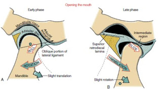
The arthrokinematics of opening the mouth are depicted for an early and a late phase in Figure 11-16. The early phase, constituting the first 35% to 50% of the range of motion,
involves primarily rotation of the mandible relative to the cranium.66,88 As depicted in Figure 11-16, A, the condyle rolls posteriorly within the concave inferior surface of the disc.
(The direction of the roll is described relative to the rotation
of a point on the ramus of the mandible.) The rolling motion swings the body of the mandible inferiorly and posteriorly.


The axis of rotation is not fixed but migrates within the vicinity of the condyles.24,60
The rolling motion of the condyle stretches the oblique portion of the lateral ligament. The increased tension in the ligament helps to initiate the late phase of the mouth’s opening.57,71
The late phase of opening the mouth consists of the final 50% to 65% of the total range of motion. This phase is marked by a gradual transition from primary rotation to primary translation. The transition can be readily appreciated by palpating the condyle of the mandible during the full opening of the mouth. During the translation the condyle and disc slide together in a forward and inferior direction
against the slope of the articular eminence (see Figure 11-16, B). At the end of opening, the axis of rotation shifts inferiorly. The exact point of the axis is difficult to define because it depends on the person’s unique rotation-to-translation ratio.
At the later phase of opening, the axis is usually below the neck of the mandible.24

Full opening of the mouth maximally stretches and pulls the disc anteriorly. The extent of the forward translation (protrusion) is limited, in part, by tension in the stretched, elastic superior retrodiscal lamina. The intermediate region of the disc translates forward while remaining between the superior
aspect of the condyle and the articular eminence. This placement of the disc maximizes joint congruency and reduces intra-articular stress.
The arthrokinematics of closing the mouth occur in the reverse order of that described for opening. 

When the mouth is fully opened and prepared to close, tension in the superior retrodiscal lamina starts to retract the disc, initiating the early translational phase of closing. The later phase is dominated
by rotation of the condyle within the concavity of the disc, terminated when contact is made between the upper and lower teeth.

viernes, 16 de febrero de 2018

The osteokinematics of the mandible

The osteokinematics of the mandible are most often described as protrusion and retrusion, lateral excursion, and depression and elevation. All of these movements occur to varying degrees during mastication. For a more detailed analysis of mandibular movements, the reader
is encouraged to consult the classic work by Posselt,61 thoroughly summarized by Okeson.

PROTRUSION AND RETRUSION
Protrusion of the mandible occurs as it translates anteriorly without significant rotation (see Figure 11-13, A). Protrusion is an important component of the mouth’s opening maximally.
Retrusion of the mandible occurs in the reverse direction (see Figure 11-13, B). Retrusion provides an important component of closing the widely opened and protruded mouth.


LATERAL EXCURSION
Lateral excursion of the mandible occurs primarily as a side-toside translation (see Figure 11-14, A). The direction (right or left) of active lateral excursion can be described as either contralateral
or ipsilateral to the side of the primary muscle action.In the adult, an average of 11 mm (about 1
2 inch) of maximal unilateral excursion is considered normal.74 Lateral excursion of the mandible is usually combined with other relatively slight translations and rotations. Normally the specific path of movement is guided by the shape of the mandibular fossa and position of the articular disc.


DEPRESSION AND ELEVATION
Depression of the mandible causes the mouth to open, a fundamental component of chewing (see Figure 11-15, A).
Maximal opening of the mouth typically occurs during actions such as yawning and singing. In the adult the mouth can be opened an average of 50mm as measured between the incisal edges of the upper and lower front teeth.2,35,74 The interincisal opening is typically large enough to fit three adult “knuckles” (proximal interphalangeal joints). Typical mastication, however, requires an average maximal opening of 18 mm—about 36% of maximum (sufficient to accept one adult knuckle). Being unable to fit two knuckles between the edges of the upper and lower incisors is usually considered abnormal in the average sized adult. Elevation of the mandible closes the mouth—an action used to grind food during mastication (see Figure 11-15, B).


TOMADO DE KINESIOLOGY OF THE  MUSCULOSKELETAL  SYSTEM .
AUTOR. DONALD NEUMANN

viernes, 9 de febrero de 2018

Importancia del juego para las personas mayores


Nuestro cerebro también necesita un poco de “gimnasia”. Hay actividades cognitivas que son tan beneficiosas para nuestra mente como lo es el ejercicio físico moderado para nuestro cuerpo.
Un estudio norteamericano de la RSNA (Sociedad Radiológica de Norteamérica) dirigido por el doctor Konstantinos Arfanakis, del Centro Médico Universitario Rush de Chicago (Illinois, EE.UU.) y realizado en personas de una media de edad de 81 años, aconseja cinco actividades concretas
para esta edad: leer, escribir, jugar al ajedrez, ir al teatro y frecuentar las bibliotecas.

Estas actividades (y otras similares, lógicamente) ayudan a preservar la  integridad estructural en el cerebro de las personas mayores. O si lo prefieren decir de una manera llana y simple, mantener el cerebro ocupado en edades avanzadas da “resultados positivos”.
Si usted quiere ponerse a hacer un poco de “gimnasia mental” y hacer trabajar su memoria, por nombrar un ejemplo de capacidad cognitiva, puede tomar el listín telefónico y empezar a memorizar nombres y números, calles, etc.
Bien, éste es un sistema sin duda un poco expeditivo y poco recomendable de poner en práctica, aunque sólo sea por el riesgo inminente de morir de aburrimiento atroz.

Pero no se preocupe, pues en estas páginas le propongo hacerlo de una manera mucho más divertida y –muy posiblemente– también más eficaz: con diversos juegos de todo tipo con los cuales, además de aprender, reforzar o potenciar sus capacidades cognitivas, podrá relacionarse con otras personas de su entorno más o menos inmediato: familiares, amigos y conocidos.
El ser humano, como todo ser vivo, está sometido a cambios que, a lo largo de los primeros años de su vida implican crecimiento y desarrollo, y que se producen muy rápidamente.
Por el contrario, en la edad adulta los cambios son mucho más lentos y en algunas de nuestras capacidades se producen declives en su rendimiento y pérdidas funcionales.


Sin embargo, como señalan los especialistas en la materia, esos declives y pérdidas pueden ser compensados. Ello se consigue optimizando el desarrollo personal y haciendo que los factores de crecimiento existan a lo largo de todo el ciclo vital, buscando oportunidades de desarrollo, mejorando potencialidades y compensando el déficit que pueda ir apareciendo.
Pensemos que, al menos en parte, la pérdida de habilidades con motivo de la edad puede deberse a la falta de uso o su progresivo o repentino abandono.
Normalmente una persona que se jubila reduce su entorno habitual, así como el número de personas con las que trata, incluso se mueve menos, va a menos actividades culturales o recreativas, etc.

Al hacer menos actividades o menos variadas disminuye sus fuentes de estimulación y sus posibilidades de enfrentarse con situaciones nuevas.
Poco a poco se va relajando y finalmente se puede acabar haciendo dependiente de los demás cuando quizás fácilmente se podría haber evitado.

Ya sólo falta que cuando se le olvida algo le digamos que eso es normal y entonces deje de esforzarse, en cuanto a actividad mental y física se refiere.
La disminución de la inteligencia que se produce en personas mayores que no tienen enfermedades, habitualmente puede ser compensada mediante entrenamiento y práctica. Sólo es necesario tener ganas y hacer un pequeño esfuerzo que sin duda pronto se verá recompensado.
Y es que las personas mayores tienen una amplia capacidad de reserva que les permite compensar la disminución de algunas de sus capacidades.
Así, la experiencia y la práctica les pueden ayudar a resolver determinados tipo de cuestiones.


Recuerdo cuando empecé a trabajar por casualidad como chocolatero –una dulce profesión que nada tenía que ver con mis estudios universitarios, con doctorado incluido– sin tener ningún conocimiento sobre el oficio, pero con ganas e ilusión por aprender. El encargado del obradoren el que trabajaba, el Sr. Eugeni, estaba a punto de jubilarse y siempre me dio la impresión que llevaba un ritmo lento y cansino en su tarea, pero nada más alejado de la realidad, pues aquella persona sabía suplir muy bien sus carencias gracias a su experiencia y saber hacer, de manera que cuando yo –llevado por mi empuje juvenil– intentaba hacer algo más deprisa que él, me resultaba del todo imposible lograrlo, pues él con su experiencia y técnica muy perfeccionada hacía de forma aparentemente fácil lo que para una persona inexperta como yo resultaba todo un reto, ya que yo no sólo tenía que hacer las cosas deprisa, sino bien, naturalmente.


Como se afirma en la Guía del Juguete (2011): “El juego potencia la autonomía de los mayores, ayuda a mantener y mejorar la salud y facilita y consolida nuestras redes y apoyo social”.
El envejecimiento no tiene que ser sinónimo de una vida sedentaria y rutinaria. Al contrario, este periodo de nuestra vida puede ser una etapa llena de vitalidad si así nos lo proponemos. Hay que romper con los falsos mitos y creencias de que envejecer nos impide hacer cosas divertidas y
sociales. Es más, el jugar y participar de actividades recreativas ofrece un marco para que las personas mayores tengan un lugar donde sociabilizarse, entretenerse y sentirse realmente vivas.
Podemos pues, afirmar con rotundidad, que los juegos en la tercera edad son muy productivos y altamente recomendados.

ARTICULO TOMADO DEL LIBRO: GIMNASIA MENTAL PARA MAYORES DE JORGE  BATLLORI

domingo, 10 de abril de 2016

SHAPE OF THE PROXIMAL FEMUR (Short paper of Newman's Kinesiology)

SHAPE OF THE PROXIMAL FEMUR

The ultimate shape and configuration of the developing proximal femur are determined by several factors, including differential growth of the bone’s ossification centers, the force of muscle activation and weight bearing, and circulation.

Abnormal growth and development resulting in a misshaped proximal femur is referred to generically as femoral dysplasia (from the Greek dys, ill or bad, + plasia, growth). Trauma or other acquired factors can also affect the shape of the proximal femur. The shape and configuration of the proximal femur have important implications on the congruity and stability of the joint, as well as the stress placed on the joint structures. This topic will be revisited throughout this chapter. Two specific angulations of the proximal femur help define its shape: the angle of inclination and the torsional angle.
Angle of Inclination The angle of inclination of the proximal femur describes the angle within the frontal plane between the femoral neck and the medial side of the femoral shaft (Figure 12-7). At birth this angle measures about 140 to 150 degrees. Primarily because of the loading across the femoral neck during walking, this angle usually reduces to its normal adulthood value of about 125 degrees.16,132 As depicted by the pair of red dots in Figure 12-7, this angle optimizes the alignment of the joint surfaces.
A change in the normal angle of inclination is referred to as either coxa vara or coxa valga. Coxa vara (Latin coxa, hip, + vara, to bend inward) describes an angle of inclination markedly less than 125 degrees; coxa valga (Latin valga, to bend outward) describes an angle of inclination markedly greater than 125 degrees (see Figure 12-7, B and C). These abnormal angles can significantly alter the articulation between the femoral head and the acetabulum, thereby affecting hip biomechanics. Severe malalignment may lead to dislocation or stress-induced degeneration of the joint.

Femoral Torsion Femoral torsion describes the relative rotation (twist) between the bone’s shaft and neck. Normally, as viewed from above, the femoral neck projects about 15 degrees anterior to a medial-lateral axis through the femoral condyles.43 This degree of torsion is called normal anteversion (Figure 12-8, A). In conjunction with the normal angle of inclination, an approximate 15-degree angle of anteversion affords optimal alignment and joint congruence (see alignment of red dots in  Figure 12-8, A).

Femoral torsion that is markedly different from 15 degrees is considered abnormal. Torsion significantly greater than 15 degrees is called excessive anteversion (see Figure 12-8, B). In contrast, torsion significantly less than 15 degrees (i.e., approaching 0 degrees) is called retroversion (see Figure 12-8, C). Typically a healthy infant is born with about 40 degrees of femoral anteversion.43 With continued bone growth, increased weight bearing, and muscle activity, this angle usually decreases to about 15 degrees by 16 years of age. Excessive anteversion that persists into adulthood can increase the likelihood of hip dislocation, articular incongruence, increased joint contact force, and increased wear on the articular cartilage.57 These factors may lead to secondary osteoarthritis of the hip.147

Excessive anteversion in children may be associated with an abnormal gait pattern called “in-toeing.” In-toeing is a walking pattern with exaggerated posturing of hip internal rotation. The amount of in-toeing is generally related to the amount of femoral anteversion. This gait pattern apparently is a compensatory mechanism used to guide the excessively anteverted femoral head more directly into the acetabulum (Figure 12-9). In addition, Arnold and colleagues have shown that the exaggerated internally rotated position during walking serves to increase the moment arm of the important hip abductor muscles—leverage that is substantially reduced with excessive femoral anteversion.9

Regardless of the reason for the internal rotated position, children may,  over time, develop shortening of the internal rotator muscles and various ligaments, thereby reducing external rotation range of motion. Fortunately, most children with in-toeing eventually walk normally.163 The gait pattern typically improves with time because of a natural normalization of the anteversion or a combined structural compensation in other parts of the lower extremity, most commonly the tibia.60


There is no evidence that nonoperative treatment can reduce excessive femoral anteversion. Excessive femoral anteversion of 25 to 45 degrees is common in persons with cerebral palsy, and even anteversion as high as 60 to 80 degrees has been reported.8,16 In-toeing typically persists in children with cerebral palsy who are ambulatory and usually does not resolve